Using AI and Ultrasound to Diagnose COVID-19 Faster

Coronavirus disease 2019 (COVID-19) is a newly identified virus that has caused a recent outbreak of respiratory illnesses starting from an isolated event to a global pandemic. As of July 2020, there are over 2.8 million confirmed COVID-19 cases in the U.S. and over 11.4 million worldwide. In the United States alone, over 130,000 Americans have died from COVID-19, with no end in sight. A major cause of this rapid and seemingly endless expansion can be traced back to the inefficiency and shortage of testing kits that offer accurate results in a timely manner. The lack of optimized tools necessary for rapid mass testing produces a ripple effect that includes the health of your loved ones, jobs, education, and on the national level, a country’s Gross Domestic Product (GDP), but artificial intelligence and ultrasound may help.

STATE OF ART IN DIAGNOSIS

Prof. Alper Yilmaz, PhDCurrently, there are two types of tests that are conducted by healthcare professionals–diagnostic tests and antibody tests. The diagnostic test, as the name implies, helps diagnose an active coronavirus infection in a patient. The ideal diagnostic test and the “gold standard” according to the United States Center for Disease Control (CDC) is the Reverse Transcription Polymerase Chain Reaction, or simply, RT-PCR. RT-PCR is a molecular test not only capable of diagnosing an active coronavirus infection, but it can also indicate whether the patient has ever had COVID-19 or were infected with the coronavirus in the past. However, the time required to conduct the test limits its effectiveness when mass deployed.

A much faster but less reliable diagnostic test alternative to RT-PCR is an antigen test. Much like the gold standard, the antigen test is capable of detecting an active coronavirus infection in a much shorter timeframe. Although antigen tests produce rapid results, usually in about an hour, the results are deemed highly unreliable, especially with patients who were tested negative according to the US FDA.

In contrast, the antibody test is designed to search for antibodies produced by the immune system of a patient in response to the virus and is limited by its ability to only detect past infections, which is less than ideal to prevent an ongoing pandemic.

THE PROBLEM 

To combat the rapid expansion of an airborne virus such as COVID-19, or future variations of a similar virus, rapid and reliable solutions must be developed that aim at improving the limitations of current methods. Although highly accurate, methods such as RT-PCR do not meet the speed requirements needed for testing on a large scale. Depending on the location, diagnosis of an active coronavirus infection with RT-PCR may take anywhere between several hours and up to a week. When the number of daily human-to-human interactions are considered, the lack of speed in diagnosing an active coronavirus patient could be the difference between a pandemic or an isolated local event.

As an alternative to molecular tests, Computed Tomography (CT) scans of a patient’s chest have shown promising results in detecting an infection. However, in addition to not being recommended by the CDC to diagnose COVID-19 patients, there are many unwanted consequences with the use of CT scans. With CT scans used to diagnose multiple illnesses, some of which relate to serious emergencies such as brain hemorrhaging, they cannot be used as the primary tool for diagnosing COVID-19. This is especially true in rural areas where the healthcare infrastructure is underfunded. Mainly due to the required deep cleaning of the machine and room after each patient, which usually requires 60 to 120 minutes, many institutions are unable to provide CT scans as a viable primary diagnostic tool. Ultimately, given the need for CT scanners for several other health complications combined with limited patient capacity at each hospital, alternative methods must be developed to diagnose an active coronavirus patient.

THE SOLUTION 

Recently Point-of-Care (POC) devices have started to be adopted by many healthcare professionals due to its reliability and portability. An emerging popular technique, which adopts improvements made in mobile ultrasound technology, allows for healthcare professionals to conduct rapid screenings on a large scale.

Working since mid-March, when early cases of physicians adopting mobile ultrasound technology emerged, the research team at The Ohio State University, Dr. Alper Yilmaz and PhD student Shehan Perera, started developing a solution that can automate an already well-established process. Dr. Yilmaz is the director of the Photogrammetric Computer Vision lab at Ohio State. Dr. Yilmaz’s expertise in machine learning, artificial intelligence, and computer vision combined with the research experience of Shehan Perera laid a strong foundation to tackle the problem at hand. As it stands, the screening of a new patient, with the use of a mobile ultrasound device takes about 13 minutes, with the caveat that it requires a highly trained professional to interpret the results generated by the device. With the combination of deep learning and computer vision, the research team was able to use data generated from the ultrasound device to accurately identify COVID-19 cases. The current network architecture, which is the product of many iterations, is capable of detecting the presence of the virus in a patient with a high level of accuracy.

Many fields have been revolutionized with modern deep learning and computer vision technologies. With the methods developed by the research team, this technology can now allow any untrained worker to use a handheld ultrasound device, and still be able to provide a service that rivals that of a highly trained doctor. In addition to being extremely accurate, the automated detection and diagnosis process takes less than 10 minutes, which includes scanning time, and sanitation is as simple as removing a plastic seal that covers the device. The benefits of this technology can not only be useful for countries such as the United States, with a well-established healthcare system, but, more importantly, can significantly help countries and areas where medical expertise is rare.

CONCLUSION 

The United States healthcare system is among the best in the world, yet we are failing to provide the necessary treatment patients clearly need. The developments made in artificial intelligence, deep learning, and computer vision offer proven benefits, which can not only be leveraged to improve the current state of the global pandemic but can lay the foundation to prevent the next. Alternative testing methods such as mobile ultrasound devices combined with novel artificial intelligence algorithms that allow for mass production, distribution, and testing could be the innovation that could help decelerate the spread of the virus, reducing the strain on the global healthcare infrastructure.

Feel Free to Reach the Authors at: 

Photogrammetric Computer Vision Lab – https://pcvlab.engineering.osu.edu/
Dr. Alper Yilmaz, PhD
Email: Yilmaz.15@osu.du
LinkedIn: https://www.linkedin.com/in/alper-yilmaz

Shehan Perera
Email: Perera.27@osu.edu
LinkedIn: https://www.linkedin.com/in/shehanp/

References 

https://www.fda.gov/consumers/consumer-updates/coronavirus-testing-basics

https://www.whitehouse.gov/articles/depth-look-COVID-19s-early-effects-consumer-spending-gdp/#:~:text=BEA%20estimates%20that%20real%20GDP,first%20decline%20in%20six%20years.&text=This%20drop%20in%20GDP%20serves,in%20response%20to%20COVID%2D19.


Interested in learning more about COVID-19 or AI? Check out the following posts from the Scan:

https://connect.aium.org/home

The Invisible Front Line

2020’s trials seem to have come on like a freight train; full steam ahead with no signs of stopping. Australia was still burning when we first heard stories of a novel virus with pandemic potential in Wuhan, China. Numbers and other details seemed to change daily. Weeks went by as we watched world news intently, taking note of the infection rate and death toll, all the while steeling ourselves for a possible outbreak at home. As much as we tried to go about our daily lives, Wuhan and the virus was never too far from our minds. Was this virus airborne? There were still so many unanswered questions, but one thing was certain; COVID-19 was spreading like wildfire and it was only a matter of time now before we would be on our own front line.Huang

Sonographers and other medical professionals soon began deployment into COVID wards in our own hospitals: areas that had been sealed off and outfitted as negative pressure cohort units to treat the infected patients. Then the deluge of daily updates and dizzying policy changes began as we tried to keep up with CDC guidelines. Rumors surfaced of limited PPE (personal protective equipment) supplies. Only doctors and nurses needed n95s? Regular procedure masks were fine for everyone else? Surely that was incorrect. Surely they knew what kind of prolonged contact sonographers have with our patients? X-ray was making contact with every patient under investigation (PUI). CT was scanning countless chests. Worries intensified as we all tried to navigate this new reality.

I’ll never forget my first assignment in the cohort. Only one other sonographer in my department had gone into the cohort at that time. He relayed seeing 3 morgue carts lining a hallway on his first trip inside. I thought about that often in the days that followed and I knew my turn was coming. How would I handle that? Some of our respiratory therapy (RT) and interventional radiology (IR) colleagues had tested positive by this time. I thought about my little boy. I saw news coverage of doctors and nurses who were self-quarantining after their shifts to decrease the potential spread to their families. I didn’t have that option as a single mother.

Finally, it came: my first COVID+ request. I told myself it would be fine. I just needed to be brave, be safe, and stay alert. I’ve never been to battle but having the media images in my mind and knowing the death toll numbers, I imagined this is what it might feel like on some small level. I thought about the PPE shortage and the rumors that we wouldn’t have access to n95s. I steeled my nerves and walked one foot in front of the other with Apollo (my LOGIQ E10). I arrived outside the cohort and was immediately greeted by the plastic sheeting that sealed off the unit. I found an anteroom with shelves overflowing with supplies. A lovely volunteer helped outfit me with everything I needed: a fresh n95, a surgical mask to go on top, a contact gown, shoe covers, eye protection, and a scrub hat. We exchanged nervous chatter for a moment as she gave me a once over to make sure I was ready. She opened the door and I exhaled as I walked inside.

As I made my way to my first patient, I noticed things were definitely different. Physicians and nurses donned full respirator masks, patient information was written on the room windows so staff could see information such as code status from the hallway, and iv poles with extra tubing sat outside of patient rooms so nurses could adjust pumps without going inside. I also learned that doctors were either doing virtual or modified rounds with one MD per team going into the patient’s room while the rest stayed outside. One came in during my 30-minute exam. As I stood hip-to-hip with my patient, he stood at the foot of the bed, asked the patient a few questions, and was gone in about 2 minutes. It struck me how much extra caution was being taken for doctors and nurses to limit their exposure times.

Some other things in the cohort looked like business as usual. I saw radiographers and cardiac sonographers going about their usual work. I saw food service delivering meals. I saw housekeeping working to stay on top of the mountains of doffed contact gowns and other garbage. Everyone was working individually on this front line for a common goal: our patients. Yet, as I arrived home that day and turned on the news, I was once again told by the media that nurses and doctors are the essential workers in this pandemic. While I absolutely believe nurses and doctors deserve every ounce of recognition they receive, I sometimes think people forget that it takes a team to deliver excellent patient care. I was fortunate enough to be able to share my experiences with Alison Bowen of the Chicago Tribune recently in the hopes of illuminating just some of what we do in a day as Diagnostic Medical Sonographers.

My first patient had a seizure during my exam that day. As I approached my second patient’s room to perform a liver Doppler, a doctor sitting outside of the room informed me the patient had just passed away. My third patient was about to receive a Foley catheter and was extremely nervous. Her nurse asked me to help assist before I started my ultrasound. The patient was still very nervous so I went to the hallway to find extra help. I asked an employee there if she wouldn’t mind coming in and holding the patient’s hand. She looked behind herself and then back at me before stating, “I’m just EVS [environmental services] but I’m happy to help if it’s OK.” She donned a gown and jumped right in.


Angela Huang, BS, RDMS (AB,OB/GYN,PS), RVT, is a Diagnostic Medical Sonographer for a large research hospital in Chicago. She attended DePaul University for undergraduate studies where she majored in Biology. Huang went on to Sonography school at El Centro College in Dallas, Texas. Now, she has a 10-year-old son who keeps her laughing and they love to travel and explore.

Interested in learning more about COVID-19? Check out the following posts from the Scan:


https://connect.aium.org/home

No Words Are Strong Enough

Low- and middle-income countries have always faced major health difficulties related to lack of human resources, facilities, and access to drinking water and electricity. Added to these factors are the lack of a suitable road, geographical remoteness, and poverty. Hence, the management of patients is compromised both diagnostically and therapeutically.

IMG_20200513_125259_B

Point-of-care ultrasound (POCUS) offers wide possibilities to health professionals who work in areas with limited resources by means of the portable machine with a good battery. Therefore it is possible for the clinician to go to low- and middle-income countries to dispense quality care services on the spot while giving access to diagnostics and guiding the management and emergency invasive procedures. IMG_20200513_100229

As a primary care physician, I was trained on clinical ultrasound through the Canadian platform in the emergency unit. I use it in my routine practice as part of my physical exam with my patients, which greatly increases my precision. No words are strong enough to describe how we feel when we examine a young woman who consults for severe pelvic pains associated with metrorrhagia and we suspect an ectopic pregnancy and the B-HCG urine test comes out positive, so you grab your US probe and you find an empty uterus, a hemoperitoneum. The fact that you saw the patient’s interior and were to be able to show her what exactly is wrong…. It’s a strength beyond what the words can explain, the precise diagnosis is reliable and prompt.

Once a month, I travel to Yabassi, a small village surrounded by a forest in the littoral region of Cameroon, which is difficult to access and rarely supplied with electricity, to do ultrasound for pregnant women discouraged by the bad state of the road and the distance to reach the nearest town. I help them meet their babies for the first time and I enable adequate follow up for the pregnancy and prevent certain complications that might occur during the delivery.

With a minimum of 1 doctor for 30,000 people, it is imperative for the clinician to go to the patients and not the reverse. And POCUS can help in these situations because of its ability to save the images to be shown to other experts for their expertise if needed. Ultrasound offers immense possibilities in upper-income countries, and I think it’s even more important in low- and middle-income countries to have access to that highly efficient and accessible method, to greatly improve the management of patients while offering quality healthcare at a low cost.


Yannick Ndefo, MD, is a general practitioner at St Thomas hospital in Douala, Cameroon.

Interested in learning more about ultrasound in low-resource settings? Check out the following posts from the Scan:

Do It With Heart: Pre-Intubation Point-of-Care Echocardiography for Hemodynamic Optimization

Have you ever wondered why that patient coded after endotracheal intubation? As it turns out, it is not uncommon after critically ill patients are intubated. Approximately 60% of critically ill patients require endotracheal intubation and are at high risk for hemodynamic collapse during this procedure. Prior studies suggest that there is up to a 25% risk of hemodynamic instability even in successful critical care unit intubations. Therefore, it is crucial to prevent peri-intubation hemodynamic instability to avoid poor patient outcomes through hemodynamic optimization prior to endotracheal intubation.

Point-of-care ultrasound has evolved as a simple, portable, and noninvasive tool for assessment of hemodynamic status. It can provide invaluable information about diagnoses and direct resuscitation in critically ill patients. This bedside imaging modality can help determine the etiology of shock, guide appropriate interventions prior to patient decompensation, and assess patient response to management changes. It can also assist in the evaluation of intravascular volume status and fluid responsiveness of critically ill patients.

Endotracheal intubation is especially perilous for a patient with right ventricular failure. Performing this procedure in patients with right ventricular failure can result in catastrophic hemodynamic collapse since the right heart is very sensitive to increases in afterload. Right ventricular failure resulting in hemodynamic collapse is an underappreciated complication of patients undergoing intubation and invasive mechanical ventilation.

Echocardiography during the preparation period of intubation allows for direct and noninvasive visualization of the right ventricle at the bedside and can play a major role in the stabilization of critically ill patients. Pre-intubation echocardiography can prevent hemodynamic deterioration by identifying a failing right ventricle, which is extremely sensitive and unable to compensate for any increase in afterload or decrease in preload from endotracheal intubation. Pre-intubation echocardiography can detect signs of a deteriorating right ventricle (pressure and volume overload) such as right ventricle dilation, bowing of the interventricular septum into the left ventricle, decrease in the size of the left ventricular cavity, and decreased left ventricular filling leading to decreased cardiac output (Figures 1–4). If acute right ventricular failure is identified prior to endotracheal intubation, it can help the physician select appropriate management strategies prior to intubation and avoid hemodynamic instability.


With pre-intubation detection of right ventricular failure, different strategies can be implemented prior to endotracheal intubation to avoid hemodynamic collapse. Non-invasive positive pressure ventilation can be an alternative in some cases, which has a less pronounced effect on venous return and preload compared to invasive mechanical ventilation. In the setting of pulmonary embolism (or pulmonary arterial hypertension), inhaled nitric oxide can be used to decrease pulmonary artery pressure through pulmonary vascular dilation. Other strategies to avoid worsening right ventricular failure include administration of vasopressors prior to endotracheal intubation and avoiding intravenous fluid boluses.

Pre-intubation echocardiography is a crucial step in the protocol during endotracheal intubation of critically ill patients to prevent poor patient outcomes. It allows clinicians to approach endotracheal intubation-associated hemodynamic instability in a specific, targeted manner. Integration of pre-intubation echocardiography can vastly improve the management and safety of critically ill patients, in hopes of decreasing the risk of poor outcomes.


Srikar Adhikari, MD, MS, FAIUM, is a professor in the Department of Emergency Medicine at the University of Arizona Medical Center.

Interested in learning more about POCUS? Check out the following posts from the Scan:


Hey, Ultrasound! What Did I Do Without You?

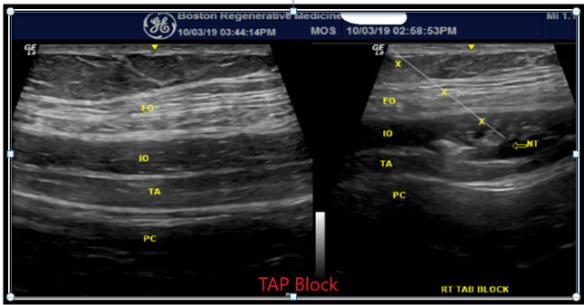
I trained as a physiatrist, which means a great deal of education on musculoskeletal conditions. Over the course of my residency training, I became more and more comfortable with bony and soft tissue landmarks for examination and targeting various joints, nerves, and tendons for therapeutic injections. As I was supervised by attendings, and carefully followed their instructions, there was no doubt in my mind that the tip of my needle was at the target intended. Why would I doubt a common practice that has been in existence for several decades?Mostoufi

As I started my fellowship in spine/pain/musculoskeletal care, I found the love of my life, the fluoroscope!!  Here, I had access to a tool that made life incredibly easy. I actually could visualize my targeted hip, shoulder, or facet joint, and inject some contrast to identify the needle tip within my target. I could precisely deliver therapeutic medications to a particular nerve root, and even identify vascular uptake and avoid procedural complications.

It was then and there that I realized that there were substantial shortcomings in what I learned as “landmark-based injections”. I realized that even though I had learned the proper “blind” procedure technique, there was no confirmation that my medication had reached its intended target. More importantly, if my patient did not respond to the procedure, I could not differentiate between a medical condition that was not responsive to the treatment versus shortcomings of un-guided procedures and inadequate delivery of medications to the targeted tissue/joint. For 12 years, I confidently treated thousands of patients by performing spine and musculoskeletal injections using my fluoroscope. I enjoyed using my C-arm, and life was pretty good.

In 2011, while attending a PM&R national conference, I sat through a 15-minute presentation on overdiagnosis of trochanteric bursitis. The speaker eloquently described fluoroscopic-guided bursa injection. This was something that I did on a regular basis as a diagnostic step. He then used ultrasound (US) images to demonstrate a few cases of gluteus medius tendinopathy and also trochanteric bursitis and how US can be superior to X-ray in therapeutic sub-gluteus maximus bursa injection. While sitting and listening, I recognized that it was virtually impossible to press against the lateral trochanter and be accurate about the diagnosis. It is also not possible to use fluoroscopy and be sure that the steroid or regenerative treatments are correctly delivered to sub-gluteus maximus bursa.

Remembering how helpful fluoroscope was to identify particular bony landmarks and assist with the proper treatment of spine and joint disease, here I was discovering a new tool that can enhance diagnostic and therapeutic skills in musculoskeletal care in particular soft tissue disease (nerves, muscles, tendons). This meant a fundamental change in the way I was going to treat patients but also a change in how I train the next generations of Physiatrists, coming through our residency program.

Fig 3aFig 3b

Learning to use the US, and incorporating it into the practice was much harder than I envisioned and also very expensive. At the time, there were limited well-structured educational resources available, and the learning curve was quite steep. As I was learning, I had to beg (or pay) my kids to become my scanning subjects!!

In contrast to a fluoroscope, it is nearly impossible to recognize an abnormal structure on the US unless you are comfortable with the normal anatomy. With a ton of hands-on workshops, mentorship, practice, and with assistance from my new found love of ultrasound machine, and guidelines from the AIUM, ultrasound has become easier and more enjoyable!! The abnormal findings became more clear and treatments more effective. In this process, I found out that patients enjoy looking at the US screen and being explained about finding on a screen full of gray, gray, and grayer lines and curves.

US has transformed how physiatrists practice and teach musculoskeletal medicine. Point-of-care US imaging allows for the residents and fellows to visualize various organs or structures within an organ, recognize healthy and diseased tissue, and diagnose the problem on the spot. This, in turn, will lead to a quick and targeted treatment and satisfied patients.

Examples of musculoskeletal (MSK) conditions that US has proven to be an effective tool to workup or treat includes rotator cuff and biceps tendinopathy, small or large joint injections, upper extremity nerve entrapments, muscle and tendon tears, peripheral nerve lesions, carpal tunnel syndrome (CTS), intersection syndromes, trigger fingers, plantar fasciitis, piriformis and sciatic complaints, treatments of bursitis or tenosynovitis, iliotibial  (IT) band treatment, ischiofemoral impingement, and many diagnoses for which dynamic testing proves to be beneficial.

Fig 6

Despite its cost and extensive training/certification needs, utilization of US in MSK care is predicted to be a standard of care in the next 5–10 years. As more and more practitioners are trained, its use for diagnostic or therapeutic purposes will become the norm.

Fig 7aFig 7b

I still love my fluoroscope and prefer its use in most spine procedures. Adding US has revolutionized my practice and allows me to be a better diagnostician, a better MSK doctor and a better educator for both my patients as well as future providers that come after me. In short, US has been a game-changer.


Ali Mostoufi, MD, FAAPMR, FAAPM, is an Assistant Prof. in PM&R at Tufts University, and the president of New England Spine Care Associates (NeSpineCare.com) and Boston Regenerative Medicine (BostonRegen.com).  As a spine and sports medicine practitioner, his clinical practice focuses on Interventional Spine, Diagnostic US, US-based therapeutic interventions and Regenerative Medicine in spine and sports.

Interested in reading more about musculoskeletal ultrasound? Check out the following posts from the Scan:


Comment below, or, AIUM members, continue the conversation on Connect, the AIUM’s online community to share your experience.

https://connect.aium.org/home


My Sonography Experience With COVID-19

It is been almost 5 weeks since I got infected with SARS-CoV-2 (also known as COVID-19), my life-changing experience.1

The day all started, during my night shift, I started with low-grade fever, chills, and myalgia; I did not doubt for a second that I had to have the test for SARS-CoV-2. That same day, most of my mild COVID-19 patients had these same cold-like symptoms, but some of them did not have a known epidemiological contact. Without time to have any other tests done, laboratory or X-ray, I self-quarantined at home waiting for the result. And finally, it came in the midst of the night; I received the “positive”.

In the morning, as more symptoms started to appear, headache, diarrhea, anosmia, ageusia and dry cough, it was a relief to have my hand-held ultrasound device at home. With the rush, I even left my oximeter, which measures heart rate and blood oxygen levels, in my hospital locker.

There is now growing evidence regarding the imaging findings of COVID-19, but at that time, the only studies were performed via CT scan and X-ray. With my ultrasound probe, I scanned following 8 zones (2 anterior, 2 lateral of both hemithorax) plus posterior lobes. I felt relieved (didn’t last long) to see there was a normal A-line pattern. More relief came when at some point I had a dull but constant right lower abdominal pain with normal appendix and no hydronephrosis on ultrasound.


What impresses most about this disease is its dynamic pattern, with sudden changes during the evolution. As my symptoms waxed and waned, so did my lung ultrasound, probably in a different manner than I would have expected. As the disease progressed, I saw all the possible lung findings, from the initial posterobasal scattered B-lines, to small pleural effusions, irregular pleural line, coalescent B-lines, and finally subpleural consolidations, especially in posterior and lateral areas. My personal impression was that I wasn’t feeling worse when I had more B-lines, but when the subpleural consolidations started to appear and spread. Each time I had new subpleural consolidations, there was a worsening in my symptoms coming: more myasthenia, cough, and diarrhea. After the second week, the subpleural consolidations were replaced by coalescent and scattered B-lines. Following that, the irregular pleural line persisted longer.

March 22 still


Surprisingly, during the third week, things started to worsen again, and on ultrasound there was a big consolidation appearing in one lobe, that was my sign for a therapy shift towards antibiotics.

My personal feeling is that consolidations are more reliable than just the number of B-lines, and correlated better with my symptoms. Actually, after 3 weeks from the symptom onset, after recovering and testing negative for SARS-CoV-2, I still had several areas with scattered and coalescent B-lines, as well as thickening of the pleural line. We have to be more flexible and take into account other parameters (i.e. oximetry), rather than rely solely on the number of affected areas on ultrasound, to compose the clinical picture, and influence the management.

As I remarked before, what impresses me most about this disease is the ultrasound dynamism. After having recovered, I still had new areas of thickening of pleural line that appeared in the back (asymptomatic) for the following week (4th), and almost 5 weeks after, I still had one plaque. And after 5 weeks, I am still surprised to have unnoticed findings, such as an asymptomatic pericardial effusion.

As a firm sonobeliever, I found it extremely useful to monitor my disease for sonographic progression and or resolution, and quickly detect complications. After this experience and having returned to work, I would have no excuse to irradiate my patients before scanning them, in the same way I went through.

Definitely, this experience was the best lesson I could have before returning to the trenches.


Yale Tung Chen, MD, PhD, is an associate professor at Universidad Alfonso X El Sabio, in Madrid, Spain. He was diagnosed with COVID-19 and shared his symptoms and ultrasound images each day on Twitter @yaletung. Follow his thread at #mycoviddiary.

Interested in reading about topics that could be of interest during the COVID-19 pandemic? Check out the following posts from the Scan:

The Personal Touch: The importance of human interactions in ultrasound

As I write this, the novel coronavirus COVID-19 is spreading across the globe, inciting fear and anxiety. Aside from frequent hand-washing and other routine precautions, many leaders, officials, and bloggers are advocating for limiting person-to-person contact. This has resulted in cancelation of many professional society meetings, sporting events, and social gatherings, and has stimulated new conversations regarding working from home and virtual meetings. Although these suggestions have many clear benefits (such as the decreased burden of commuting; limiting the spread of infection), there are additional reports describing the impact loss of face-to-face interactions may have on job satisfaction, workflow efficiency, and quality.Fetzer-David-14-2

The current practice of medicine, more than ever, relies on a team approach. No one individual has the time, knowledge, or experience to tackle all aspects of an individual’s care. No one is an island. Unlike many television shows that highlight a single physician performing everything from brain surgery to infectious disease testing, the reality is that we each rely on countless other members of the healthcare team. That practice of medical imaging, ultrasound, in particular, is no different. Whether we work in a radiology, cardiology or vascular, or obstetrical/gynecology practice, the team, and more importantly the relationship between team members, is paramount to an effective and impactful practice.

As a radiologist in a busy academic center, I rely on and value my personal relationship with my team of 50+ sonographers. These relationships have been facilitated by day-to-day, face-to-face interactions, allowing me to get to know the person behind the ultrasound images. These interactions foster an environment of trust. For my most experienced sonographers, my implicit trust ultimately leads to fast, efficient and precise exam interpretations, while for sonographers I rarely work with, my index of suspicion regarding a finding is naturally heightened, impacting my confidence in my diagnosis and thus affecting my interpretation, and ultimately how my report drives patient care.

The trust goes both ways: a strong relationship also fosters honest communication whereby sonographers can come to me with questions or concerns regarding exam appropriateness, adjustments to imaging protocols, and the relevance of a specific imaging finding. The direct interaction provides an opportunity for sonographers, new and experienced, to be provided immediate direct feedback regarding their study—they can learn from me, and often I from them, making us all that much better at the end of the workday.

In addition to trust, open communication allows for users of ultrasound to take advantage of one of the key differentiating features of ultrasound compared to other modalities: the dynamic, real-time nature of image acquisition. Protocol variations can be discussed on-the-fly. Preliminary findings can be shared with the interpreter, and additional images can be obtained immediately, without having to rely on call-backs, inaccurate reports, and reliance of follow up imaging (often by other modalities). This ultimately enhances patient care and decreases healthcare costs. In our practice, we have the ability to add contrast-enhanced ultrasound for an incidental finding, allowing us to make definitive diagnoses immediately, without having to recommend a CT or MRI—this would not be possible if it were not for a personalized checkout process.

We continue to hear about changes in ultrasound workflow across the country: sonographers and physicians, small groups and large, academic and private practices have all considered or have already implemented changes that minimize the communication between sonographer and study interpreter. This places more responsibility on the sonographer to function independently, and minimizes or even eliminates the opportunities for quality control and education. Sonographer notes and worksheets, and electronic QA systems, are poor substitutes for the often more nuanced human interaction. In my experience, these personal encounters enhance job satisfaction, and the lack of it risks stagnating learning and personal drive. There have been many sonographers that have left local practices to join our medical center specifically to take advantage of the sonographer-radiologist interaction we continue to nurture.

Some elements driving these transformations are difficult to change: growing numbers of patients; increasing reliance on medical imaging; medical group consolidation; etc. Many changes to sonographer workflow have been fueled by a focus on efficiency (decreasing scan time, improving modality turn-around times, etc.). Unfortunately, these changes have been made with little regard to how limiting team member communication impacts examination quality, job satisfaction, and patient outcomes; for those of you in a position to address workflow changes, consider these factors. For sonographers yearning for this relationship, do not be afraid to reach out to your colleagues and supervising physicians—ask questions, be curious, and engage with them. Nearly everyone appreciates a human interaction, and even the toughest personality can be cracked with a smile and some persistence. In the end, it is the human interactions and the open and honest communication that not only make us better healthcare providers but happier and healthier human beings.


David Fetzer, MD, is an assistant professor in the Abdominal Imaging Division, as well as is the Medical Director of Ultrasound in the Department of Radiology at the UT Southwestern Medical Center.


Interested in reading more about communication? Check out the following posts from the Scan:

The Best of the Scan, 5 Years in the Making

The Scan has been a home for all things ultrasound, from accreditation to zoos, since its debut 5 years ago, on February 6, 2015.MISC_SCAN_5_YR_ANN_DIGITAL_ASSETS_FB

In its first 5 years, the Scan has seen exponential growth, in large part due to the hard work of our 110 writers, who have volunteered their time to provide the 134 posts that are available on this anniversary. And it all began with Why Not Start? by Peter Magnuson, the AIUM’s Director of Communications and Member Services, who spearheaded the blog’s development.

In honor of this 5th Anniversary, here are some of your favorites:

Top 5 Most Viewed Posts

unnamed

1. Ultrasound Can Catch What NIPT Misses
by Simcha Yagel
(August 4, 2015)

Sonographer Stretches2. Sonographer Stretches for an ‘A’ Game
by Doug Wuebben and Mark Roozen
(January 31, 2017)

Keepsake3. The Issue with Keepsake Ultrasounds
by Peter Magnuson
(April 30, 2015)

Hip Flexor Stretch4. 3 Stretches All Sonographers Should Do
by Doug Wuebben and Mark Roozen
(January 19, 2016)

Anton5. From Sonographer to Ultrasound Practitioner: My Career Journey
by Tracy Anton
(October 23, 2018)

The Fastest Growing Posts
That Are Not Already in the Top 5

And we have plenty more great posts, such as:

POCUS: A Holiday in the Sun

Getting started with point-of-care ultrasound (POCUS) is like taking a vacation in Bali, Bermuda, or the Bahamas.  Let’s say you’ve landed in an exotic destination and plan to rent a car to explore the island. After collecting your keys, what’s next? Jump in the vehicle and peel off to the beach? Of course not – you’ll take a minute to consider the controls of your car, where you’re going, and how you’ll get there. POCUS is no different from a dream island visit.

Mackenzie headshot 2

In an unfamiliar vehicle, it’s normal to become acquainted with the controls. You want to know how to turn on the car’s lights and wipers and position the mirrors and windows appropriately. There’s a direct analog in performing a POCUS study. The operator has to select the correct transducer and examination preset before getting started. If it’s a machine you’re not familiar with, you need to take a moment to locate essential controls such as depth and gain. Even if the machine is familiar, you need to optimize those settings to ensure you can obtain quality images, just as you would with the mirrors in your car.

It’s also second-hand nature to adjust a car for comfort. The seats and steering wheel need to be positioned so you have a comfortable trip, and the climate settings arranged for passenger comfort. For a successful POCUS scan, the same steps should happen. Both the operator and the patient should be comfortable and positioned correctly. That means adjusting the bed, lowering the side rails, and placing the patient and machine where you can obtain adequate images while ensuring no one has to be a contortionist.

Taking a car on the road on unfamiliar roads can be stressful, and more so if you’re not used to driving on the left. If driving on the opposite side of the road is unfamiliar, it’s smart to visualize how you will be oriented on the road and during turns before heading out on the road. Successful POCUS users have the same habit: they understand where the indicator marker is on both the screen and the transducer before acquiring images. Failing to do so leads to confusion and a breakdown of pattern recognition, just as driving on the left might.

With the car and orientation controls sorted, you’re still not going to fire up the engine yet. Most travelers take a moment to figure out where they’re headed, with a GPS or map. The sonologist needs to take the same step, remembering the focused question they’re trying to answer with the POCUS study, and what they need to see to be satisfied. While you might be happy to ramble aimlessly in a car, POCUS scans should stay focused.

Of course, this assumes that renting a car is the best way to get around the island. Maybe you’d be better served by a taxi, bus, or boat. Or maybe after seeing the rental vehicle, you decide the car can’t accommodate your plans. In the same vein, not all clinical questions can be answered with POCUS. An alternative imaging modality or comprehensive ultrasound may be the test you need, and it’s OK to change your mind and decide you need something else after you perform the scan.

POCUS is rewarding and helps both clinicians and patients, but isn’t always easy. Think of POCUS like the start of a vacation, and you may find your studies are easier, and a bit closer to a holiday in the sun.


Comment below, or, AIUM members, continue the conversation on Connect, the AIUM’s online community to share your experience.

Connect_digital_graphics_E-NEWSLETTER

David Mackenzie, MDCM, is an emergency physician at Maine Medical Center, in Portland, Maine. Follow Dr Mackenzie on Twitter @mackendc.

Novice to Competence to Understanding Our Role as POCUS Educators

Nights at the VA medical ICU could get lonely sometimes. When the hubbub of the day had drawn down and the critical care fellows had gone home, the work in the ICUs slowed.Headshot_kevin piro

I figured that I would make use of the time that had seemingly stopped. I grabbed the ultrasound and went to scan and chat with a friendly gentleman whom I had admitted the previous night. It became readily apparent that I was still a struggling learner at this point in my training. There was something that looked like cardiac motion, but not resembling anything like the diagrams and videos I had looked at on my own. It was an uncomfortable place to be.

I imagine that is where a lot of people get frustrated and stop, especially when they don’t have someone to encourage and nurture their continued practice. I had a different luxury. Just a few weeks prior, I had received an inquiry about participating in a new general medicine POCUS fellowship at Oregon Health & Science University, and I was instantly sold on its potential. Here was a chance to carve out a new path and to invest in a skill that offered me a skillset that could improve my patient care. And I knew that I would have the benefit of POCUS experts literally holding my hand as I learned the skill. What a luxury!

So, I kept scanning in the ICU prior to my fellowship. You know what I found? Patients are much more forgiving than we might imagine them to be. Most understand that hospitals are frequently places of learning and like to be engaged in the process and, as I stumbled through my next few exams, I was reminded of my Dad’s words of encouragement, “the only difference between you and an expert is that they have done it once or twice.” So I kept at it. I was terrible the next times too. But, it got easier and I felt less intimated with each scan I performed. By the time I hit fellowship, I was already moving in the right direction.

When I started my POCUS fellowship, I was fortunate to work with all sorts of supportive colleagues that allowed me to continue to grow. Where I had struggled to build a foundation on my own, colleagues collected from internists, sonographers, and EM physicians provided me with the scaffolding. They provided me with lessons. “Remember, air is the enemy of ultrasound” and “ultrasound does not give you permission to turn your brain off. It is a problem-solving tool.” They entertained clinical application questions. They gave back when I leaned in. These colleagues were an amazing support network and would help me construct the mosaic that I teach from now.

A few months into the fellowship, I could complete a competent exam comfortably. It came together one day for me when I completed a Cardiovascular Limited Ultrasound Exam (CLUE) on a pleasantly demented older man, who had shortness of breath likely representing heart failure. As I looked at his lungs, taking stock of the bilateral B-lines and pleural effusions that confirmed his diagnosis, I discussed and showed the findings with his daughter.

“This makes so much sense now!” she remarked. The lightbulb went on for her as I democratized her father’s clinical information. The lightbulb came on for me too as I had a sense of satisfaction of both feeling confident in my diagnosis, but also being better able to teach and engage a family in their medical care. My transformation from novice to competency was mostly complete.

Now, a little more than 2 years removed from my fellowship, I have a little more perspective on the road from novice to competency, not only from my personal experience but also from my opportunity to network with an amazing group of enthusiastic (IM) POCUS educators.

These educators are largely trained by their own curiosity, their attendance at POCUS CME courses, or by latching onto experts from peripheral medical departments. In essence, these educators are pulling themselves up by their own bootstraps in a time when there is a distinct scarcity of POCUS educators within Internal Medicine, which can leave the supposed “all-knowledgeable” physician in an uncomfortable place of vulnerability. They have shared the angst that POCUS is a particularly challenging skill to learn due to its humbling nature – we may not know how badly we were hearing murmurs as medical students, but I bet most learners can guess by looking at a picture how poorly they are doing when they are scanning. It was a feeling I shared back in the ICU as a resident, but our experiences diverged when I had mentors who invested in me learning this valuable skill.

But, these physicians who learned POCUS independently are now at the next, even harder, part. As new leaders, we must reach behind us and pull up the trainees, whether that be by creating the next POCUS fellowship, starting or improving a residency POCUS program, or simply training your fellow colleague. We are tasked with making new learners feel supported and encouraged, and to make this technology accessible in fields where POCUS is not the standard of care. We need to foster these learners’ growth so that they can arrive at their own lightbulb moment and so they keep scanning on the ICUs in the effort to improve the care they deliver.


What was your defining moment in your decision to go into ultrasound? Have you had a unique learning experience? Comment below, or, AIUM members, continue the conversation on Connect, the AIUM’s online community to share your experience.

Connect_digital_graphics_E-NEWSLETTER


Kevin M. Piro, MD, participated in and helped build a point-of-care ultrasound fellowship at Oregon Health & Science University (OHSU), becoming only the second general medicine-focused ultrasound fellowship in the nation. Dr Piro is now a hospitalist at OHSU.