Ultrasound: A Diagnostic Tool and for Treating Injuries and Diseases

Now, more than ever, staying up to date on the latest trends and innovations in ultrasound is essential for physicians. This year, the annual meeting of the American Institute of Ultrasound in Medicine (AIUM) is being transformed into UltraCon! This new conference puts you at the center of the conversation where expertise meets interaction and debate. So how can you ensure that you are prepared to take full advantage of this transformative opportunity? 

In a previous post, we highlighted how you could explore new, exciting, and current technologies in ultrasound; identify the different approaches to diagnostic ultrasound; and determine which ultrasound techniques can help you advance your practice at “Can You Do That With Ultrasound?” on day one at UltraCon (March 25–29, 2023). Day two at UltraCon offers attendees more opportunities to deepen their understanding of ultrasound—both as a diagnostic tool and in treating injuries and diseases—through two additional symposiums: “Optimizing Ultrasound Image Quality” and “Ultrasound Diagnoses You Can’t Miss.”

Optimizing Ultrasound Image Quality

Optimizing ultrasound image quality and, ultimately, patient care is at the heart of this symposium. As a healthcare professional, you will be well-equipped to succeed at this task with knowledge gained through key topics such as physics, knobology, and Doppler, as well as improving patient/probe position. You will benefit from roundtable discussions that cover image reviews and quality assurance—useful topics that can help move one step closer to improving patient outcomes. Earn 5 CMEs for learning about ultrasound technology advancements, advancing your image acquisition techniques, and finding ways to improve image quality. This symposium is sure to contribute to providing better patient care!

This symposium will feature a total of 8 engaging and interactive sessions for attendees to participate through in groups:

  • Physics: So Easy it Hertz,” led by Frederick Kremkau, PhD, FAIUM. How does ultrasound work? It’s all about physics. Knowing the role physics plays will help you avoid artifacts.
  • “Know Your Knobs,” led by Elena S. Sinkovskaya, MD, PhD. To even get an ultrasound image, you must know how your machine works, how to make fundamental adjustments, and how to make optimal use of B-mode. Glossary handout included.
  • “Elements of Scanning,” led by Margaret R. Lewis, MD. Improve your ultrasound diagnostics by understanding optimal patient and transducer positioning techniques, equipment quality assurance, and more.
  • “Demystifying Doppler,” led by Tracy Anton, MD. What is Doppler ultrasound? How does it work? How do I select the correct equipment? How do I interpret the results? Learn all this and more by attending.
  • “Just Images Roundtable,” led by Mishella Perez, BS, RDMS, RDCS, FAIUM, and Yvette S. Groszmann, MD, MPH. Learn what you can do to improve image quality across specialties, including OB, GYN, MSK, POCUS, and Vascular.
  • “Echoes of the Past to the Voices of the Future,” led by Frederick Kremkau, PhD, FAIUM. Attend this session to understand how prior advancements in ultrasound technology have established the scaffold for the possibilities of the use of diagnostic ultrasound in the present and the future.
  • “Ultrasound Quality Assurance Roundtable,” led by Timothy Canavan, MD, MSc, FAIUM, Therese Cooper, BS, RDMS, David Jones, MD, FAIUM, Anita Moon-Grady, MD, and Aubrey Rybyinski, MD. Understanding the role of accreditation and continuous QA is essential to ensuring the best outcomes for patients. Hear from a panel of experts to better guide your practice and get answers to your questions.
  • Image Quality Trivia: Test your ultrasound knowledge during this fun, quiz-style game where the entire audience participates. Topics include OB, fetal echo, GYN, physics, MSK, and general imaging.

Ultrasound Diagnoses You Can’t Miss

Ultrasound diagnoses are a crucial part of maintaining top-level medical care. Ultrasound experts from various disciplines meet in this symposium to share their expert knowledge and experience, allowing participants to stay on top of the imaging findings and avoid any form of misdiagnosis. During the symposium, a multispecialty expert panel will create an interactive discussion for the attendees to apply their learning to real clinical scenarios. And what’s more? You can earn up to 5.75 CMEs just by attending this “Ultrasound Diagnoses You Can’t Miss” symposium!

This symposium will feature a total of 17 engaging and interactive sessions for attendees to participate in where subspecialty discussion, breakout opportunities, and rapid case reviews will take place.

UltraCon is an incredible opportunity for medical ultrasound practitioners who want to stay up-to-date on all things related to medical ultrasound technology. From interactive debates to resource handouts, exhibitor sandboxes, and focus groups—this event has something for everyone! Come prepared with an open mind and get ready to connect with experts from around the world while learning about the exciting new possibilities that are transforming the field of medical ultrasound today! Don’t miss out—All of this is just what is available on the second day of symposia at UltraCon. Check out the Full Schedule to get a sneak peek at everything you could learn.

Arian Tyler, BS, is the Digital Media and Communications Coordinator for the American Institute of Ultrasound in Medicine (AIUM).

Dermatologic Ultrasound: Skin Deep Knowledge

According to the recent European Federation of Societies for Ultrasound in Medicine and Biology (EFSUMB) Position Statement on Dermatologic Ultrasound,1 dermatologic ultrasound is the application of ultrasound to the diagnosis of skin and appendages (hair and nails) diseases, as well as their healthy state, and cosmetic alterations.

This application was born as a necessity to answer challenges in dermatology that could not be addressed with superficial exploration methods such as clinical inspection, wood lamp, or dermoscopy, which mainly offer 2D views of the skin. The possibility of adding a third dimension (depth) and even a 4th one (with Doppler or dynamic explorations) with ultrasound makes dermatologic ultrasound a new way to go from guessing what’s happening below the surface to “seeing” what is really happening.

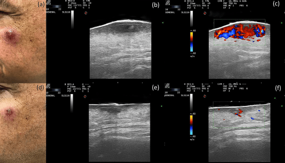
Ultrasound follow-up of intralesional glucantime injection in a cutaneous leishmaniasis. a) Clinical before treatment, b) B-mode before treatment, c) Color Doppler before treatment, d) Clinical after 3 injections, d) B-mode after 3 injections, d) Color Doppler after 3 injections.

During my years of clinical practice incorporating ultrasound in dermatology, I have heard some questions that always seem to arise in meetings. These are some of them:

What are the technical requirements for quality dermatologic ultrasound?

Although equipment with 70 MHz can be helpful to elucidate very concrete aspects of skin pathology, most questions and clinical situations in dermatology can be addressed today with high-quality conventional equipment above 15 MHz with color Doppler.

Apart from high-quality equipment, having a deep knowledge of dermatology, basic ultrasound, and its physical principles is the key to performing appropriate explorations in dermatologic ultrasound.

Which are the main fields of dermatologic ultrasound?

The fields at the forefront of dermatologic ultrasound are skin oncology, inflammatory skin diseases, and cosmetic dermatology.

How can dermatologic ultrasound help us dermatologist in skin oncology?

Skin cancer is the most frequent cancer in the human body. Most malignant skin tumor prognosis and treatment rely on depth and structural invasion, therefore, dermatologic ultrasound is an essential tool compared with MRI or CT in these kinds of evaluations, mainly because of its higher resolution and availability.

Is dermatologic ultrasound helpful in inflammatory skin diseases?

Deep inflammatory skin diseases such as hidradenitis suppurativa and morphea and sclerosing diseases usually do not have clear superficial expression. Dermatologic ultrasound is a key tool for in-depth evaluation of the inflammatory state of these diseases and is a guide for treatment and follow-up.

What are the main applications of dermatologic ultrasound in cosmetic dermatology?

Detection of dermal fillers and the complications and adverse reactions of these medical devices is essential in the management of a responsible cosmetic dermatology practice. Dermatologic ultrasound can also be an excellent tool for skin aging evaluation and anti-aging treatment evaluation.

Who is the AIUM and how can I learn Dermatologic Ultrasound from the AIUM?

The American Institute of Ultrasound in Medicine (AIUM) is the only scientific society that hosts a very active Dermatologic Ultrasound Community, which was funded in 2015 by Ximena Wortsman together with Orlando Catalano (present vice-chair of the community), Fernando Alfageme (present chair of the community and author of this post), and Claudia Gonzalez (secretary).

This community and their members are very active in AIUM meetings, hosting several scientific and didactic sessions, and has also produced some reference papers in dermatologic ultrasound.

The AIUM has hosted several sessions and workshops in AIUM meetings in the last 5 years with the collaboration of the dermatologic ultrasound community, although a need for structured teaching and learning is still necessary for those who are interested in this subspecialty.

Dermatology is an amazing application for ultrasound and it will be key in the near future for high quality, personalized skin medicine to foster the best, responsible care for our patients.


Fernando Alfageme, MD, is a Dermatologist and Chair of the Dermatologic Ultrasound Community (AIUM) as well as Codirector of the Ultrasound Learning Centre (Dermatology) at EFSUMB.

Reference

Alfageme F, Wortsman X, Catalano O, et al. European Federation of Societies for Ultrasound in Medicine and Biology (EFSUMB) Position Statement on Dermatologic Ultrasound. Ultraschall Med. 2020 May 7. Online ahead of print. doi: 10.1055/a-1161-8872.

Pioneering Ultrasound Units

If you think your ultrasound machine is out-dated, imagine if you still had to use these from as long ago as the 1940s. 

1940s

Ultrasonic Locator
Dr G. D. Ludwig, a pioneer in medical ultrasound, concentrated on the use of ultrasound to detect gallstones and other foreign bodies embedded in tissues. During his service at the Naval Medical Medical Research Institute in Bethesda, Maryland, Dr Ludwig developed this approach that is similar to the detection of flaws in metal. This is A-mode in its operation and was Dr Ludwig’s first ultrasonic scanning equipment.

Locator


1950s

Ultrasonic Cardioscope
Designed and built by the University of Colorado Experimental Unit, the Cardioscope was intended for cardiac work.

Ultrasonic Cardioscope


1960s

Sperry Reflectoscope Pulser / Receive Unit 10N
This is an example of the first instrument to use an electronic interval counter to make axial length measurements of the eye. Individual gates for the anterior segment, lens, and vitreous compartment provided accurate measurement at 10 and 15 MHz of the axial length of the eye. This concept was the forerunner of all optical axis measurements of the eye, which are required for calculation of the appropriate intraocular lens implant power after cataract extraction. This instrument, which includes A-mode and M-mode, was developed by Dr D. Jackson Coleman and Dr Benson Carlin at the Department of Ophthalmology, Columbia Presbyterian Medical Center.

Sperry Reflectoscope Pulser


Sonoray Model No. 12 Ultrasonic Animal Tester (Branson Instruments, Inc.)
This is an intensity-modulated B-mode unit designed exclusively for animal evaluations. The instrument is housed in a rugged aluminum case with a detachable cover that contains the cables and transducer during transportation. The movable transducer holder on a fixed-curve guide was a forerunner of mechanical B-scan ultrasonic equipment.

Sonoray Animal Tester


Smith-Kline Fetal Doptone
In 1966, pharmaceutical manufacturer Smith Kline and French Laboratories of Philadelphia built and marketed a Doppler instrument called the Doptone, which was used to detect and monitor fetal blood flow and the heart rate. This instrument used the continuous wave Doppler prototype that was developed at the University of Washington. 

Smith Kline Fetal Doptone


Smith-Kline Ekoline 20
Working in collaboration with Branson Instruments of Stamford, Connecticut, Smith-Kline introduced the Ekoline 20, an A-mode and B-mode instrument for echoencephalography, in 1963. When B-mode was converted to M-mode in 1965, the Ekoline 20 became the dominant instrument for echocardiography as well as was the first instrument available for many start-up clinical diagnostic ultrasound laboratories. The A-mode was used in ophthalmology and neurology to determine brain midlines.

Ekoline 20


University of Colorado Experimental System
Developed by Douglas Howry and his team at the University of Colorado Medical Center, this compound immersion scanner included a large water-filled tank. The transducer moved back and forth along a 4-inch path while the carriage on which the transducer was mounted moved in a circle around the tank, producing secondary motion necessary for compound scanning. 

Compound immersion scannerCompound immersion scanner tub


1970s

Cromemco Z-2 Computer System (Bioengineering at the University of Washington)
This color-Doppler prototype, introduced in 1977, was the computer used for early color Doppler experiments. Z2 “microcomputers” were used for a variety of data acquisition and analysis applications, including planning combat missions for the United States Air Force and modeling braking profiles for the San Francisco Bay Area Rapid Transit (BART) system during actual operation.

Cromemco Z-2 Computer System


ADR-Model 2130
ADR of Tempe, Arizona, began delivering ultrasound components to major equipment manufacturers in 1973. Linear array real-time scanners, which began to be manufactured in the mid-1970s, provided greater resolution and more applications. Grayscale, with at least 10 shades of gray, allowed closely related soft tissues to be better differentiated. This 2-dimensional (2D) imaging machine was widely used in obstetrics and other internal medicine applications. It was marketed as an electronic linear array, which was faster and more repeatable without the need for a water bath as the transducer was placed right on the skin.

ADR Model 2130


Sonometrics Systems Inc, NY BR-400V
The first commercially available ophthalmic B-scanner, this system provided both linear and sector B-scans of the eye. The patient was examined in a water bath created around the eye by use of a sterile plastic ophthalmic drape with a central opening. Both A-scan and B-scan evaluations were possible with manual alignment of the transducer in the water bath. The instrument was developed at the Department of Ophthalmology, Columbia Presbyterian Medical Center by Dr D. Jackson Coleman, working with Frederic L. Lizzi and Louis Katz at the Riverside Research Institute.

Sonometrics Systems Inc, NY BR-400V


Unirad GZD Model 849
Unirad’s static B-scanner, allowing black-and-white anatomic imaging, was used with a scan arm and had similar controls as those used today, including processing, attenuation compensation, and gain.

Unirad GZD Model 849


1980s

American Flight Echocardiograph
This American Flight Echocardiograph (AFE) is a 43-pound off-the-shelf version of an ATL 400 medical ultrasonic imaging system, which was then modified for space shuttle compatibility by engineers at the Johnson Space Center to study the adaptations of the cardiovascular system in weightlessness. Its first journey to space was on the space shuttle Discovery in 1985 and its last on the Endeavour in 1992. The AFE generated a 2D cross-sectional image of the heart and other soft tissues and displayed it in video format at 30 frames per second. Below, Dr Fred Kremkau explains more about it.


To check out even more old ultrasound machines, visit the American Institute of Ultrasound in Medicine’s (AIUM’s) An Exhibit of Historical Ultrasound Equipment.


How old is the ultrasound machine you use now? What older ultrasound equipment have you used? Did it spark your desire to work with ultrasound? Comment below, or, AIUM members, continue the conversation on Connect, the AIUM’s online community.

Connect_digital_graphics_E-NEWSLETTER

The AIUM is a multi-disciplinary network of nearly 10,000 professionals who are committed to advancing the safe and effective use of ultrasound in medicine.

Ultrasound-Guided Cancer Imaging: The Future of Targeted Cancer Treatment

Tumor margins and malignant grade are best defined by vascular imaging modalities such as Doppler flow or contrast enhancement combined with videomicroscopy. The following are image-guided treatment options that can be performed on breast, prostate, liver, and skin cancers.

NEW DOPPLER APPLICATIONS

Blood vessel mapping using the various Doppler modalities is routinely used in both cancer treatment and reconstructive planning. In cancer surgery, it is critical to locate aberrant veins or arterial feeders in the operative site so postoperative blood loss is minimized. Advanced 3D Doppler systems allow for histogram vessel density measurement of neoplastic angiogenesis.

VESSEL DENSITY INDEX

(Fig 1) Baseline neovascularity is a treatment surrogate endpoint and therapy is maintained, increased, or suspended based on quantitative angiogenesis data.

SOLID ORGAN CANCER IMAGING UPDATES

Breast cancer, invading the lower dermis and nipple, discovered with high-resolution probes signifies the tumor has outflanked clinical observation essential for detecting the newly discovered entity of breast implant-associated anaplastic large cell lymphoma (BIA-ALCL). This capability is also vital for diagnosing the recent epidemic of male breast cancers arising near the mammographically difficult nipple areolar complex, occurring in our 911 First Responders.

For prostate cancer, 4D ultrasound can identify low-grade cancer delimited by the capsule and with low vessel density, and should be followed serially at 6-month intervals.

CONTRAST-ENHANCED ULTRASOUND (CEUS)

In 1990, Dr. Rodolfo Campani developed ultrasound contrast for liver imaging and Drs. Cosgrove (London) and Lassau (Paris) extended the use to breast, skin, and prostate tumors. CEUS is currently used worldwide but is not Food and Drug Administration (FDA)-approved in the United States.

One use for CEUS is microbubble neovascularity, which demonstrates therapeutic response since the Response Evaluation Criteria in Solid Tumors (RECIST) studies noted tumor enlargement during treatment might be related to cell death with cystic degeneration or immune cell infiltration destroying malignant tissue. Doppler ultrasound or CEUS reliably verifies decreased angiogenesis in place of contrast CT or dynamic contrast-enhanced (DCE) MRI. If vascular perfusion ceases, thermal treatments, such as cryotherapy, high-intensity focused ultrasound (HIFU), or laser ablation, should be completed.

Four-dimensional (4D) ultrasound imaging is real-time evaluation of a 3D volume so we can show the patient immediately the depth and the probability of recurrence. Specific echoes in skin cancer generated by nests of keratin are strong indicators of aggression and analyzed volumetrically. Highly suspect areas are checked for locoregional spread and a search is performed for lymphadenopathy so we can determine if the disease is confined and whether further surgical intervention is unlikely at this time. Patients are reassured because they simultaneously see the exam proceed in systematic stages. In serious cases, the patient is forewarned that the operation involves skin grafts and tissue construction.  4D ultrasound permits image-guided biopsy of the most virulent area of the dermal tumor and allows the pathologist to focus on the most suspicious region of the lymph node mass excised from the armpit, neck, or groin. Some laboratories are using postop radiography and sonography for better specimen analysis.

VIDEO DIGITAL MICROSCOPY VS BIOPSY

Fear of complications can deter patients from seeking medical opinion and surgical intervention, so many opt for noninvasive options. Imaging can help to reduce unnecessary biopsies because it can help identify the 1 out of every 33,000 moles that is malignant, while weeding out those that are not.

Once skin cancer is diagnosed, the treatment depends on depth penetration, possibly involving facial nerves, muscles around the eye and nasal bone or ear cartilage. Verified superficial tumors are treated topically or by low dose non-scarring radiation. Many cancers provoke a benign local immune response or coexistent inflammatory reaction that simulates a much larger area of malignancy, and cicatrix accompanies the healing response. 4D imaging combined with optical microscopy (RCM (reflectance confocal microscopy) or OCT (optical coherence tomography)) defines the true border during surgery, sparing healthy tissue, resulting in smaller excisional margins and less scar formation.


Do you have any tips on incorporating ultrasound in cancer imaging? Comment below, or, AIUM members, continue the conversation on Connect, the AIUM’s online community.

connect_now_live_digital_graphics_e-newsletter-1

Robert Bard, MD, DABR, FASLMS, currently runs a private consulting practice in New York City. He authored Image Guided Dermatologic Treatments, Image Guided Prostate Cancer Treatment, and DCE-MRI of Prostate Cancer and is a member of multiple leading international imaging societies. Since 1972, Dr. Bard has pioneered digital imaging technologies as alternatives to surgical biopsies for dermatologic and solid organ neoplastic disease.